
Table of Contents
Why Does the Dentist Need X‑Rays Before Treatment?
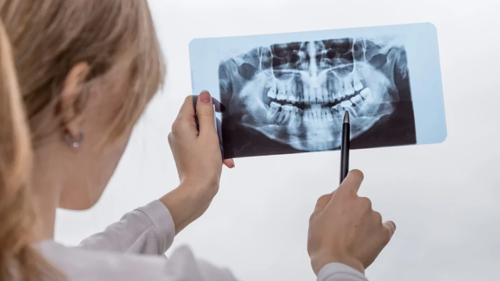
When you sit in the dental chair, you may be surprised when your dentist asks for dental X‑rays before a filling, extraction, or root canal. You might wonder: isn’t a visual exam enough? In reality, what we see in the mouth is only part of the story. Dental X‑rays show what is happening inside the tooth, under the gums, and in the jawbone – areas your dentist simply cannot see with the naked eye.
Dentists request dental X‑rays because they help them:
- Confirm a diagnosis accurately instead of relying on guesswork.
- Detect problems at an early stage, before they cause severe pain or complications.
- Choose the most appropriate treatment plan for each tooth and each patient.
- Determine root length, nerve status, and how much supporting bone is around the tooth.
- Assess treatment risks in advance, such as roots that are close to nerves or the sinus.
In other words, dental X‑rays act as a detailed map of your teeth and jaws. They allow the dentist to work more safely, reduce surprises during treatment, and improve the long‑term success of the procedures. Needing an X‑ray does not mean your case is necessarily serious; it is often a preventive step to make sure your treatment is well‑planned and suited to your individual situation.

What Do Dental X‑Rays Show?
Dental X‑rays let your dentist see below the gum line, inside the tooth, and in the surrounding bone – everything that cannot be seen directly during a regular clinical examination. With these images, many conditions can be picked up earlier, a more precise treatment plan can be made, and some painful or complex complications can be avoided later on.
Dental X‑rays can help identify:
- Hidden tooth decay
- Infections at the tooth roots
- Impacted wisdom teeth
- Jawbone loss
- Dental cysts and abscesses
- Tooth position before braces or dental implants
Hidden Tooth Decay
X‑rays are especially helpful for spotting decay between teeth or underneath existing fillings and crowns. These areas are difficult or impossible to assess fully with a mirror and probe alone. On an X‑ray, decay usually appears as a darker area compared to the rest of the tooth, which helps show how deep it is and how close it is to the nerve.
This information helps your dentist to:
- Decide whether the tooth only needs a simple filling or if root canal treatment is more appropriate.
- Identify early decay before it causes pain or leads to a tooth fracture.
- Monitor the condition of fillings and crowns over time.
Root Infections
Infections at the roots of teeth are not always obvious from the outside, and some patients may not feel constant pain. On X‑rays, an infection often shows up as a dark area or “void” around the tip of the root.
With this information, the dentist can:
- Confirm whether the tooth needs root canal treatment or retreatment.
- Evaluate how far the infection has spread into the surrounding bone.
- Track how the bone is healing after root canal treatment is completed.
Impacted Wisdom Teeth
Wisdom teeth can be tilted, partially erupted, or completely trapped in the jawbone without appearing in the mouth at all. X‑rays show:
- The direction the wisdom tooth is growing and how deeply it is buried in the bone.
- How close its roots are to the main nerve in the lower jaw or to the maxillary sinus in the upper jaw.
- Whether there are cysts or infections around an impacted wisdom tooth.
These details are important for deciding whether extraction is necessary, and if so, how to perform it with the lowest possible risk of complications. The exact approach can vary widely from one person to another.
Jawbone Loss
Healthy jawbone is essential for stable teeth and for dental implants. On X‑rays, the bone that supports the tooth roots becomes visible, allowing the dentist to assess:
- The degree of bone loss caused by long‑standing gum (periodontal) disease.
- How the bone is distributed and how high it is in different parts of the jaw.
- The severity of the problem and whether it may require specialist periodontal care or surgery.
The earlier bone loss is discovered, the easier it usually is to slow the disease and help you keep your teeth for as long as possible. Response to treatment, however, can vary from one patient to another.
Cysts and Abscesses
Some cysts or abscesses grow silently for a long time and cause little or no pain in the early stages. Dental X‑rays help to:
- Detect cysts and abscesses in the bone around tooth roots or near impacted wisdom teeth.
- Determine their size, borders, and how close they are to neighboring teeth.
- Monitor whether they are shrinking with treatment or whether surgical removal is advisable.
Finding these issues early can reduce the chance that they grow larger or affect nerves or nearby teeth. The type and extent of treatment, however, depend on the size of the lesion and on the patient’s overall health.
Tooth and Bone Position Before Braces or Implants
Before orthodontic treatment or placing dental implants, the dentist often needs a two‑ or three‑dimensional view of the teeth and surrounding bone. Dental X‑rays make it possible to:
- Assess the number, shape, and tilt of the roots before fitting braces or extracting teeth.
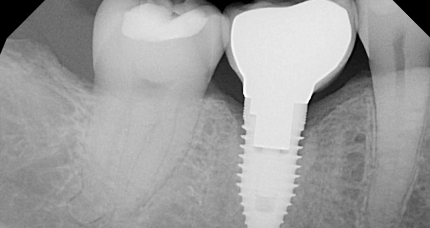
- Measure available bone for dental implants and choose the best position and angulation for each implant.
- Detect extra or unerupted teeth that might interfere with tooth movement during orthodontic treatment.
These findings help the dentist design a realistic and safe treatment plan and explain the available options to you. The suitability of any treatment type can differ from person to person and should always be discussed with your dentist.
What Types of Dental X‑Rays Are Used?
Dentists use several types of dental X‑rays and select the most appropriate one based on the problem they suspect, the planned treatment, and the patient’s age. In most clinics, dental X‑rays fall into three main categories that complement each other – no single type is used for all situations.
The main types are:
- Intraoral X‑rays
- Panoramic X‑rays
- 3D cone beam CT (CBCT)
Intraoral X‑Rays
Intraoral X‑rays are the most common type of dental X‑ray. A small film or digital sensor is placed inside the mouth near the tooth or teeth being imaged. The two most common types are periapical and bitewing X‑rays.
When Are They Used?
- To detect decay between teeth or under fillings.
- To assess tooth roots before and after root canal treatment.
- To monitor infections around tooth roots.
Advantages
- Very high detail for one or two specific teeth.
- Relatively low radiation dose compared with other types.
Your dentist chooses this type of X‑ray when they need to examine a limited area in great detail rather than the whole jaw.
Panoramic X‑Rays
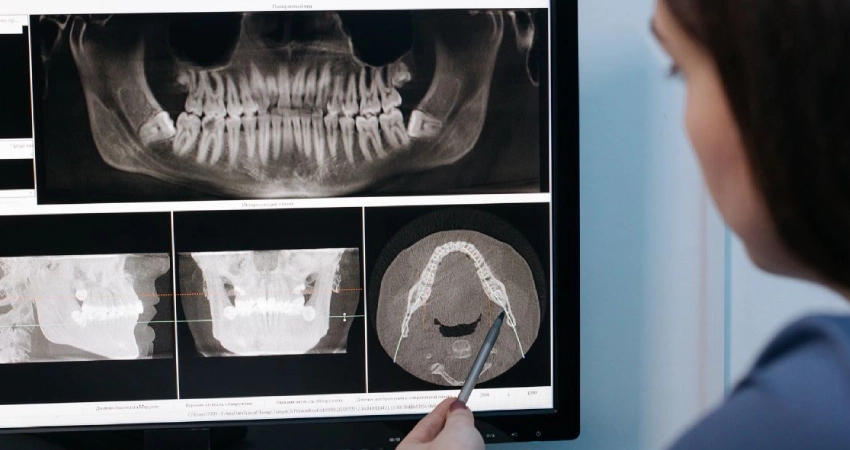
A panoramic X‑ray shows the upper and lower jaws, the temporomandibular joints (TMJ), and the maxillary sinuses in a single broad image. Nothing is placed inside your mouth; instead, the machine rotates around your head.
When Are They Used?
- To evaluate wisdom teeth and their relationship to nerves and bone.
- To check the number and position of baby and permanent teeth in children and teenagers.
- For initial planning of orthodontic treatment, implants, or oral surgery.
- To identify general jawbone problems or larger cysts and lesions.
Advantages
- Provides a quick, overall view of the jaws and teeth.
- Very useful for forming an initial treatment plan before moving on to more detailed imaging if needed.
3D Cone Beam CT (CBCT)
Cone beam CT (CBCT) is an advanced type of dental imaging that produces a 3D view of the jaws and surrounding tissues with very high detail. The images can be viewed in slices from different angles.
When Is It Used?
- For precise planning of dental implants (measuring jawbone height and thickness and locating important nerves and sinuses).
- To assess wisdom teeth that are very close to the main nerve in the lower jaw before surgery.
- To help diagnose complex cysts, tumors, or fractures in the jaws.
- In complex orthodontic cases or jaw deformities.
Advantages
- Gives highly detailed information about bone, tooth roots, and their directions.
- Helps the dentist reduce risks during complex surgical procedures.
Not every patient needs CBCT imaging. It is reserved for situations where there is a clear medical reason. Your dentist weighs the expected benefit against the additional radiation exposure and decides individually for each case.
Are Dental X‑Rays Safe?
Dental X‑rays are a routine part of modern dental care. Most international health authorities consider them safe when they are taken with modern equipment and carefully controlled doses. The amount of radiation from dental X‑rays is usually very low and significantly less than what a person is naturally exposed to from the environment over the course of a year.
Your dentist aims to keep your exposure as low as reasonably achievable and will only request X‑rays when they provide real value for diagnosis or treatment planning, not as a mere formality.
Several factors make dental X‑rays relatively safe:
- Use of modern digital systems that require less radiation than older devices.
- Protective lead aprons to shield the rest of the body and, when appropriate, a thyroid collar.
- Limiting the number and type of images to what is medically necessary, while considering the patient’s age and medical history.
If you are pregnant or planning a pregnancy, it is important to tell your dentist. In many situations, X‑rays can be postponed, or extra precautions can be taken if imaging is essential for treatment that cannot safely be delayed. The decision is made case by case, in discussion between you and your dentist.
In summary, dental X‑rays are generally considered safe when taken by a qualified dentist using approved equipment and protocols. You are always entitled to ask why a particular X‑ray is being requested and how it will help in your specific case so that you can share in the decision.
Does Every Patient Need X‑Rays Before Treatment?
Not every patient needs dental X‑rays at every visit. However, many treatments simply cannot be carried out safely and accurately without them. The decision to order X‑rays depends on your current condition, the type of problem you have, your dental and medical history, and your age.
Dentists commonly request X‑rays in situations such as:
- A first visit to a new dentist or after a long gap in check‑ups.
- Unclear pain, swelling, or other symptoms with no obvious cause.
- Planning root canal treatment or tooth extraction, especially for wisdom teeth.
- Before major treatments such as dental implants, orthodontics, or fixed bridges and crowns.
- Monitoring gum disease and bone loss around teeth.
You may not need new X‑rays if:
- You already have recent, good‑quality X‑rays of the same area.
- Your problem is simple and clearly visible during the clinical exam, such as a very shallow surface cavity.
- You attend regularly and there are no new complaints or noticeable changes.
How often you need dental X‑rays varies from person to person. Many dentists are comfortable with periodic X‑rays every one to three years for patients at lower risk of problems, while those who are more prone to decay or gum disease may need imaging more frequently. The final decision should follow a professional assessment of your situation and a discussion between you and your dentist about why X‑rays are recommended and how they may help.
How Does Dr. Ameer Ghayath Al‑Zahrawi Help You Choose the Right X‑Rays?
Choosing the right type of dental X‑ray is not random. It is based on an accurate diagnosis and a carefully considered treatment plan. Your dentist’s role is to balance the real clinical need for imaging with the goal of minimizing radiation exposure, taking into account your age and general health.
At the clinic of Dr. Ameer Ghayath Al‑Zahrawi, clear steps are typically followed, including:
1. Thorough Assessment Before Ordering Any X‑Ray
Dr. Ameer begins with a detailed medical and dental history and a full clinical examination of your teeth, gums, and mouth. If a diagnosis and treatment plan can be reached without X‑rays, additional imaging may not be requested. If there is suspicion of deep decay, root infection, a wisdom tooth problem, or preparation for implants or orthodontics, he then considers whether X‑rays are necessary and which type is most appropriate.
2. Selecting the Least and Most Accurate Imaging for Your Case
More than one type of dental X‑ray is only requested when there is a clear medical reason. For example:
- Small intraoral X‑rays to diagnose decay or root infections in a specific tooth.
- A panoramic X‑ray when a general view of wisdom teeth or jawbone is needed.
- 3D CBCT imaging only in cases that truly require high precision, such as implant planning or complex surgery.
The aim is to obtain the maximum useful information with the lowest reasonable radiation dose.
3. Explaining the Results in Simple, Clear Language
After the X‑rays are taken, Dr. Ameer explains what appears on the images: where the problem is, how severe it looks, and how it affects your teeth, gums, and bone. Seeing the issue on the screen yourself often makes it easier to understand your condition and to make an informed decision, instead of relying on a verbal description alone.
4. Linking X‑Rays to a Complete Treatment Plan
X‑rays are not a goal in themselves but a tool within a wider treatment strategy. Dr. Ameer uses them to help determine, for example:
- Whether a tooth can be treated with a filling or if root canal therapy is more suitable.
- Whether a wisdom tooth can be removed easily or if a more advanced surgical approach is needed.
- The best position for a dental implant and the thickness and height of the available bone.
The treatment plan may differ between patients even when the X‑rays look similar, depending on age, general health conditions, and individual expectations.
5. Prioritizing Safety and Minimizing Radiation Exposure
Dr. Ameer follows international safety standards, including using lead aprons when indicated and adjusting X‑ray machines to the lowest dose that still provides a clear image, especially in children and pregnant patients. In some cases, imaging may be postponed when it is considered safer to do so.
By following these steps, Dr. Ameer Ghayath Al‑Zahrawi helps you benefit from dental X‑rays as a key tool for accurate diagnosis and sound treatment planning, while protecting your safety and avoiding unnecessary imaging. The final decision is always based on open discussion between you and your dentist.